Artículos similares a Grabado de anatomía con esqueleto, cráneo y órganos de T Bromme, hacia 1865
¿Quieres más imágenes o vídeos?
Solicita imágenes o vídeos adicionales al vendedor
1 de 10
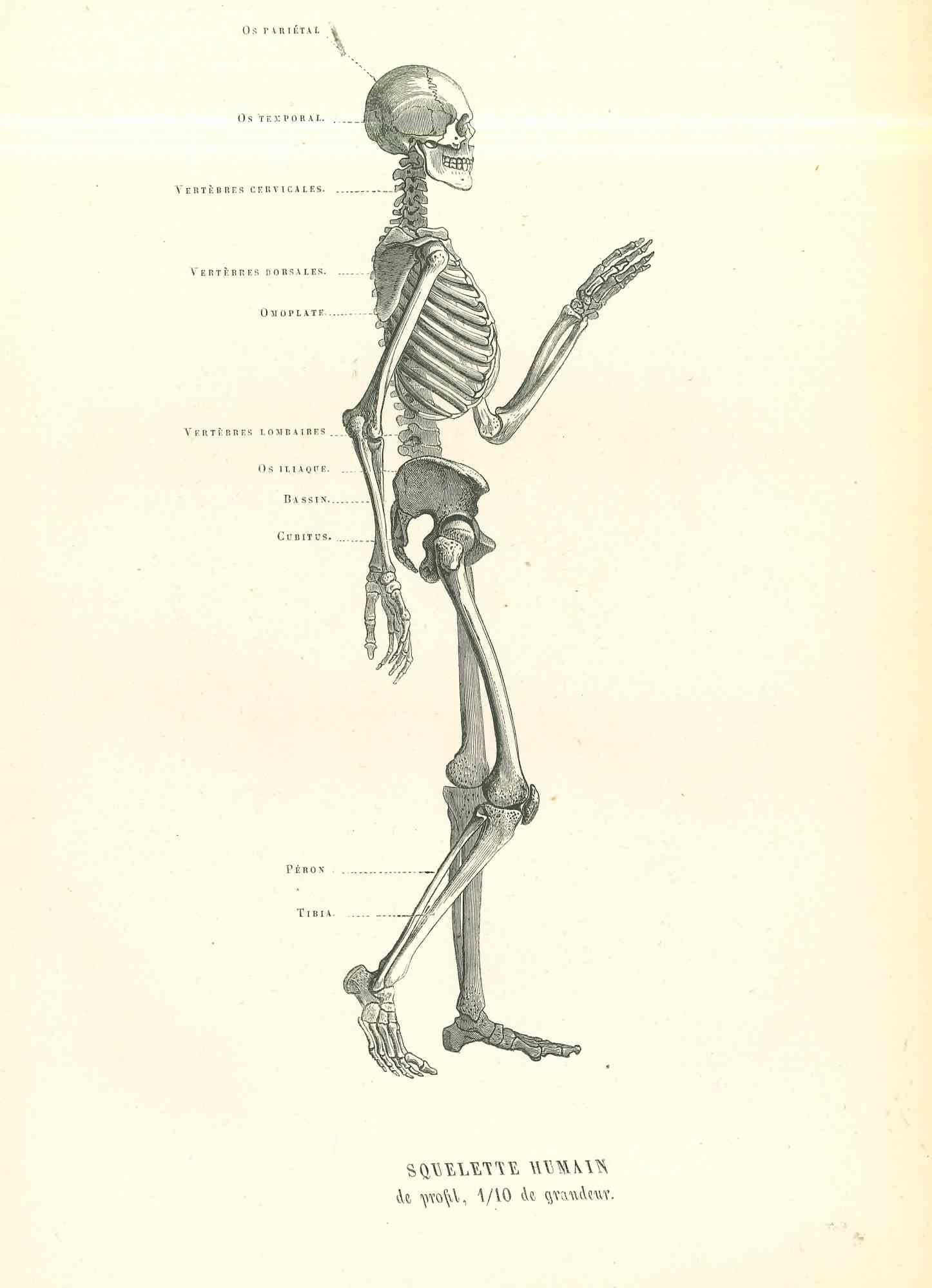
Grabado de anatomía con esqueleto, cráneo y órganos de T Bromme, hacia 1865
130 €IVA incluido
Acerca del artículo
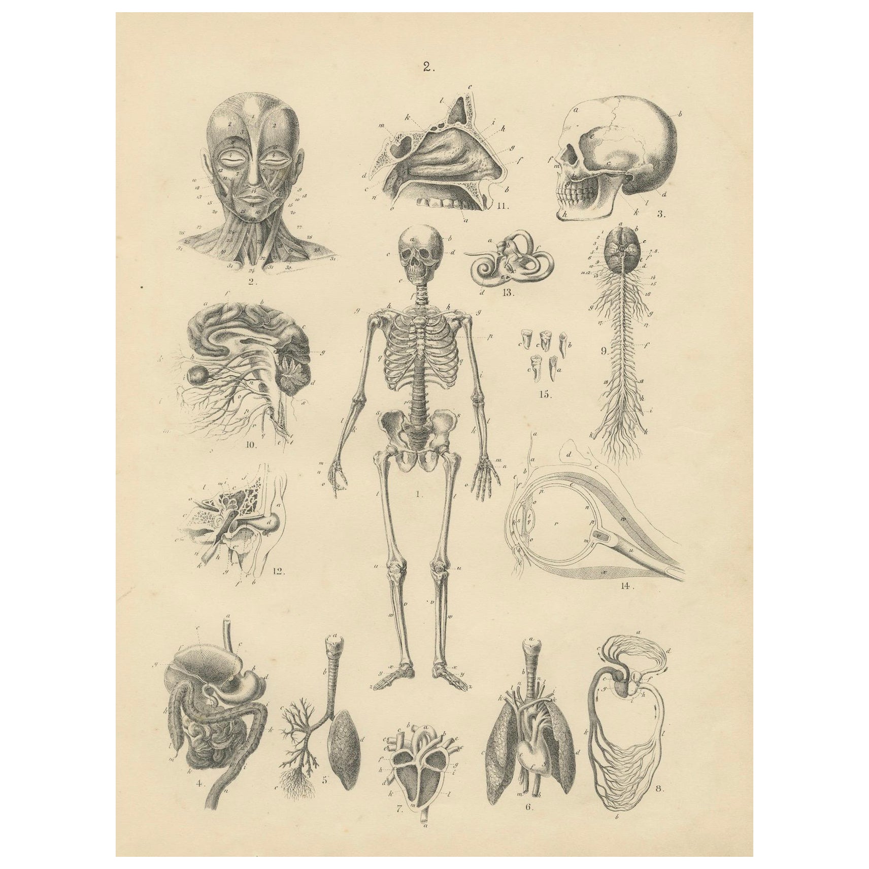
Anatomy Print with Skeleton, Skull and Organs by T Bromme, circa 1865
This original 19th-century anatomical lithograph is attributed to the German geographer and publisher Traugott Bromme and was most likely published in one of his scientific or geographic atlases around 1865. The plate is a detailed study of the human body and internal structure, featuring finely engraved illustrations of the skeleton, skull, musculature, nervous system, and various internal organs.
At the center is a full human skeleton, surrounded by smaller diagrams including the muscular face, internal organs such as the heart, lungs, stomach, intestines, brain, eyes, and inner ear. Each element is labeled with corresponding letters, indicating its original use in educational or reference contexts. This kind of diagram would have served as a visual aid for schools, medical instruction, or general scientific literacy during a time when public interest in anatomy and biology was growing alongside the rise of natural sciences.
Here is a breakdown of the numbered anatomical illustrations from the print you uploaded. Each number corresponds to a specific body part or system, based on typical 19th-century anatomical charts and their visual representation:
1. Full human skeleton
2. Muscles of the face and neck (anterior view of the facial musculature)
3. Human skull (lateral view)
4. Digestive system (esophagus, stomach, intestines)
5. Lung with bronchial branches (right lung, external structure)
6. Heart and lungs (with arteries and trachea)
7. Heart (anterior cross-section showing chambers and valves)
8. Stomach and intestines (upper digestive tract with internal folds)
9. Spinal cord and brain stem (nervous system, including spinal nerves)
10. Brain with cranial nerves (lateral view showing cerebrum and nerves)
11. Nasal cavity and sinus anatomy (sagittal section)
12. Kidney and adrenal gland with renal arteries and veins
13. Inner ear (cochlea and semicircular canals)
14. Human eye (horizontal cross-section showing retina, lens, and optic nerve)
15. Teeth and jawbones (molars, incisors, and jaw sections)
This style of diagram was intended for educational use, so the lettered parts within each figure likely correspond to detailed Latin labels in the original publication.
Traugott Bromme was known for creating illustrated atlases that encompassed not just geography, but also zoology, ethnography, and human anatomy. His works are prized today for their visual clarity and historical significance, offering a rare glimpse into how 19th-century Europeans conceptualized and taught the structure of the human body.
The lithographic technique used here displays meticulous line work, creating depth and clarity in each anatomical rendering. This print would make a valuable addition to any collection focused on medical history, early science, or antique prints.
Condition report:
Original 19th-century lithograph in good condition with light age toning and minimal edge wear. No tears or folds. Strong impression with crisp detail throughout.
Tips for framing:
Frame with a neutral-toned mat such as ivory or light grey to highlight the precision of the engraving. A black or dark wood frame adds contrast and lends the piece a classical, academic presentation suitable for display in libraries, offices, or medical settings.
- Dimensiones:Altura: 28,5 cm (11,23 in)Anchura: 22 cm (8,67 in)Profundidad: 0,2 mm (0,01 in)
- Materiales y técnicas:
- Época:
- Fecha de fabricación:1865
- Estado:Original 19th-century lithograph in good condition with light age toning and minimal edge wear. No tears or folds. Strong impression with crisp detail throughout.
- Ubicación del vendedor:Langweer, NL
- Número de referencia:Vendedor: BG-12980-21stDibs: LU3054345340142
Sobre el vendedor
5,0
Vendedor reconocido
Estos prestigiosos vendedores son líderes del sector y representan el escalón más alto en cuanto a calidad y diseño de artículos.
Vendedor Platino
Vendedores premium con una calificación de +4,7 y tiempos de respuesta de 24 horas
Establecido en 2009
Vendedor de 1stDibs desde 2017
2513 ventas en 1stDibs
Tiempo de respuesta usual: <1 hora
- EnvíoRecuperando presupuesto…Envío desde: Langweer, Países Bajos
- Política de devolución
Partes de esta página se han traducido automáticamente. 1stDibs no puede garantizar la exactitud de las traducciones. El inglés es el idioma predeterminado de este sitio web.
Garantía de autenticidad
En el improbable caso de que haya algún problema con la autenticidad de un artículo, ponte en contacto con nosotros en un plazo de 1 año para recibir un reembolso total. DetallesGarantía de devolución de dinero
Si tu artículo no es como se describe, sufre daños durante el transporte o no llega, ponte en contacto con nosotros en un plazo de 7 días para recibir un reembolso total. DetallesCancelación dentro de las 24 horas
Tienes un período de gracia de 24 horas para reconsiderar tu compra, sin preguntas.Vendedores profesionales aprobados
Nuestros vendedores de primera clase deben cumplir estrictos estándares de servicio para mantener la integridad de nuestros anuncios.Garantía de igualación de precios
Si encuentras que un vendedor publicó el mismo artículo por un precio menor en otro lado, igualaremos ese precio.Entrega global de confianza
Nuestra red de transporte de primera ofrece opciones de envío especializado en todo el mundo, que incluye envío personalizado.Más de este vendedor
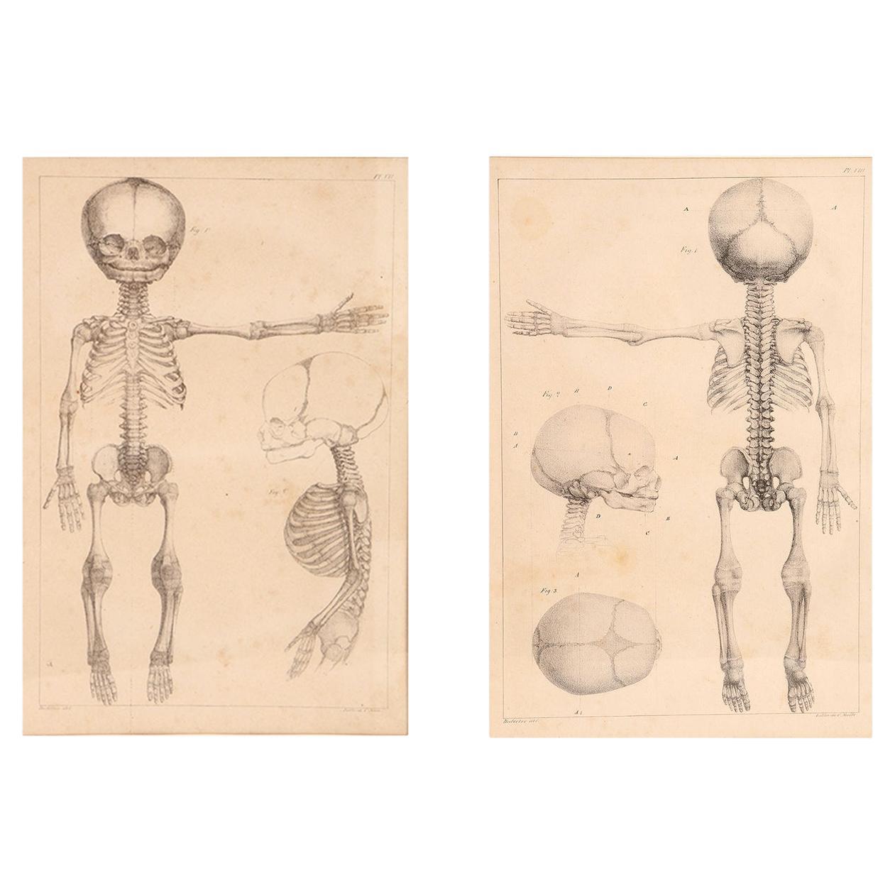
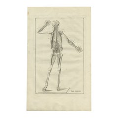
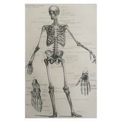
Ver todoGrabado de Anatomía: Estudio del Esqueleto, Cráneo, Órganos y Nervios - Antigüedad 1867
Grabado de Anatomía: Estudio del Esqueleto, Cráneo, Órganos y Nervios - Antigüedad 1867
Este detallado grabado antiguo presenta un completo estudio anatómico del siglo XIX, que repr...
Categoría
Antiguo, Década de 1860, Alemán, Impresiones
Materiales
Papel
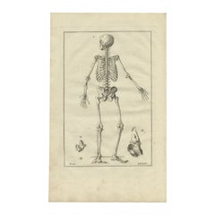
Grabado antiguo decorativo de anatomía del esqueleto humano, 1798
Grabado antiguo de anatomía del esqueleto humano. Este grabado procede de "De Ontleedkundige Plaaten van B. Eustachius", publicado por J. B. Elwe.
Artistas y grabadores: Bartolome...
Categoría
Antiguo, siglo XVIII, Impresiones
Materiales
Papel
272 € Precio de venta
Descuento del 20 %
Grabado antiguo de anatomía del esqueleto humano, 1798
Grabado antiguo de anatomía del esqueleto humano. Este grabado procede de "De Ontleedkundige Plaaten van B. Eustachius", publicado por J. B. Elwe.
Artistas y grabadores: Bartolome...
Categoría
Antiguo, siglo XVIII, Impresiones
Materiales
Papel
272 € Precio de venta
Descuento del 20 %
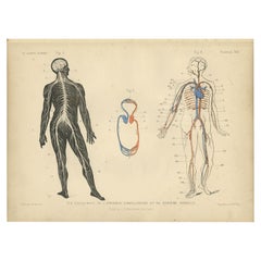
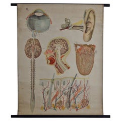
Grabado antiguo del Sistema Circulatorio y el Sistema Nervioso, 1879
Grabado antiguo titulado 'Vista de conjunto del aparato circulatorio y del sistema nervioso'. Litografía en color del sistema circulatorio y el sistema nervioso. Este grabado procede...
Categoría
Antiguo, siglo XIX, Impresiones
Materiales
Papel
128 € Precio de venta
Descuento del 20 %
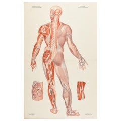
Grabado Anatómico Antiguo del Sistema Nervioso en el Cuerpo Humano '1843'
Placa anatómica coloreada (nº 17), que muestra el sistema nervioso en el cuerpo humano. De la "Anatomía elemental" de Jean-Baptiste Bourgery, publicada en 1843, ilustrada por Nicolas...
Categoría
Antiguo, Mediados del siglo XIX, Impresiones
Materiales
Papel
900 € Precio de venta
Descuento del 25 %
También te puede gustar
Grabado anatómico sobre papel, que representa un esqueleto de feto, Francia, Siglo XIX
Rara pareja de litografías anatómicas francesas del esqueleto del feto, en marco metálico de época. Gran calidad y composición artística.
Impreso por Charles Motte. París, principi...
Categoría
Antiguo, principios del siglo XIX, Francés, Impresiones
Materiales
Metal

Original Vintage Medical Print-Esqueleto, circa 1900
Gran imagen de interés médico.
Sin enmarcar.
Publicado, hacia 1900.
Categoría
Antiguo, Principios del 1900, Inglés, Eduardiano, Impresiones
Materiales
Papel
Esqueleto anatómico humano - Litografía original de Paul Gervais - 1854
Por Paul Gervais
Esqueleto anatómico humano es una litografía original sobre papel color marfil, realizada por Paul Gervais (1816-1879). La obra pertenece a la serie de "Les Trois Règnes de la Nature...
Categoría
Década de 1850, Moderno, Impresiones figurativas
Materiales
Litografía
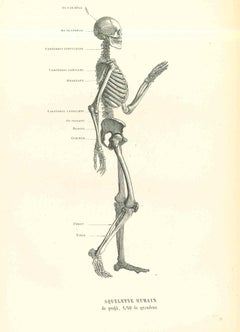
168 € Precio de venta
Descuento del 40 %
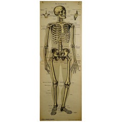
Antiguo Cuadro Anatómico de Pared que Representa el Esqueleto Humano
Raro gráfico anatómico mural del siglo XIX que representa el esqueleto humano. En los laterales Hay una descripción detallada en alemán. El gráfico mural está impreso en cartón, publ...
Categoría
principios del siglo XX, Alemán, Victoriano, Impresiones
Materiales
Papel
Antiguo Anatómico Arquitectura de la Anatomía Humana por E. Hoelemann
Una gran tabla anatómica antigua que representa los diferentes órganos de los sentidos. la tabla es la hoja número VI como parte de la arquitectura de la anatomía humana publicada po...
Categoría
Antiguo, Principios del 1900, Escandinavo moderno, Carteles
Materiales
Papel
Dibujo anatómico de un esqueleto, realizado a lápiz y tinta, Italia 1890.
Cuadro que contiene un antiguo dibujo anatómico a lápiz y tinta de un esqueleto, creado para la confección de tablas anatómicas. El marco antiguo, de madera de abeto con esquinas red...
Categoría
Antiguo, Fines del siglo XIX, Italiano, Pinturas
Materiales
Hierro
