Items Similar to Anatomy Print with Skeleton, Skull and Organs by T Bromme, circa 1865
Want more images or videos?
Request additional images or videos from the seller
1 of 10
Anatomy Print with Skeleton, Skull and Organs by T Bromme, circa 1865
$156.13
£114.99
€130
CA$212.40
A$236.02
CHF 123.68
MX$2,894.41
NOK 1,577.45
SEK 1,484.01
DKK 989.67
Shipping
Retrieving quote...The 1stDibs Promise:
Authenticity Guarantee,
Money-Back Guarantee,
24-Hour Cancellation
About the Item
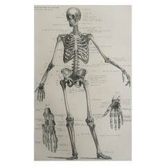
Anatomy Print with Skeleton, Skull and Organs by T Bromme, circa 1865
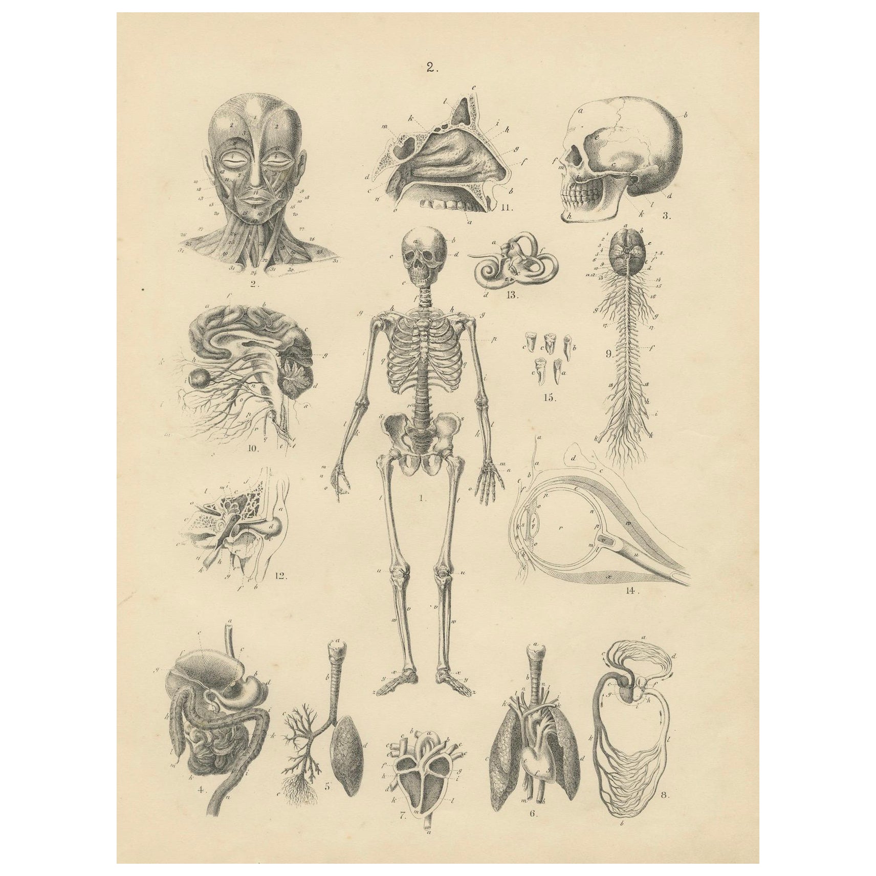
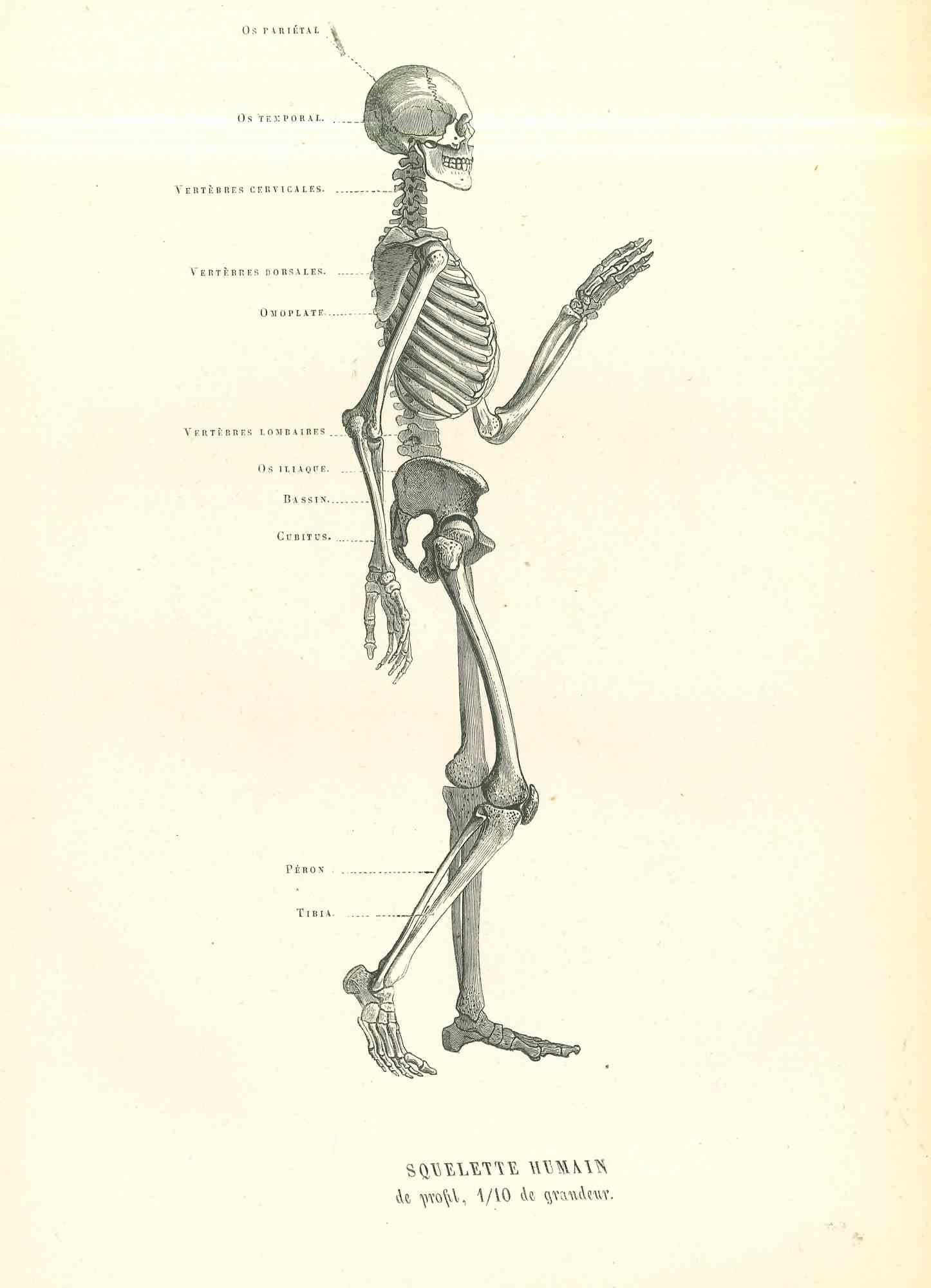
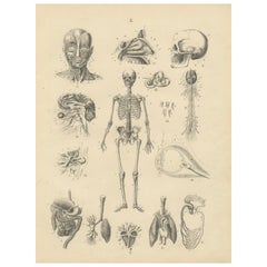
This original 19th-century anatomical lithograph is attributed to the German geographer and publisher Traugott Bromme and was most likely published in one of his scientific or geographic atlases around 1865. The plate is a detailed study of the human body and internal structure, featuring finely engraved illustrations of the skeleton, skull, musculature, nervous system, and various internal organs.
At the center is a full human skeleton, surrounded by smaller diagrams including the muscular face, internal organs such as the heart, lungs, stomach, intestines, brain, eyes, and inner ear. Each element is labeled with corresponding letters, indicating its original use in educational or reference contexts. This kind of diagram would have served as a visual aid for schools, medical instruction, or general scientific literacy during a time when public interest in anatomy and biology was growing alongside the rise of natural sciences.
Here is a breakdown of the numbered anatomical illustrations from the print you uploaded. Each number corresponds to a specific body part or system, based on typical 19th-century anatomical charts and their visual representation:
1. Full human skeleton
2. Muscles of the face and neck (anterior view of the facial musculature)
3. Human skull (lateral view)
4. Digestive system (esophagus, stomach, intestines)
5. Lung with bronchial branches (right lung, external structure)
6. Heart and lungs (with arteries and trachea)
7. Heart (anterior cross-section showing chambers and valves)
8. Stomach and intestines (upper digestive tract with internal folds)
9. Spinal cord and brain stem (nervous system, including spinal nerves)
10. Brain with cranial nerves (lateral view showing cerebrum and nerves)
11. Nasal cavity and sinus anatomy (sagittal section)
12. Kidney and adrenal gland with renal arteries and veins
13. Inner ear (cochlea and semicircular canals)
14. Human eye (horizontal cross-section showing retina, lens, and optic nerve)
15. Teeth and jawbones (molars, incisors, and jaw sections)
This style of diagram was intended for educational use, so the lettered parts within each figure likely correspond to detailed Latin labels in the original publication.
Traugott Bromme was known for creating illustrated atlases that encompassed not just geography, but also zoology, ethnography, and human anatomy. His works are prized today for their visual clarity and historical significance, offering a rare glimpse into how 19th-century Europeans conceptualized and taught the structure of the human body.
The lithographic technique used here displays meticulous line work, creating depth and clarity in each anatomical rendering. This print would make a valuable addition to any collection focused on medical history, early science, or antique prints.
Condition report:
Original 19th-century lithograph in good condition with light age toning and minimal edge wear. No tears or folds. Strong impression with crisp detail throughout.
Tips for framing:
Frame with a neutral-toned mat such as ivory or light grey to highlight the precision of the engraving. A black or dark wood frame adds contrast and lends the piece a classical, academic presentation suitable for display in libraries, offices, or medical settings.
- Dimensions:Height: 11.23 in (28.5 cm)Width: 8.67 in (22 cm)Depth: 0.01 in (0.2 mm)
- Materials and Techniques:
- Period:
- Date of Manufacture:1865
- Condition:Original 19th-century lithograph in good condition with light age toning and minimal edge wear. No tears or folds. Strong impression with crisp detail throughout.
- Seller Location:Langweer, NL
- Reference Number:Seller: BG-12980-21stDibs: LU3054345340142
About the Seller
5.0
Recognized Seller
These prestigious sellers are industry leaders and represent the highest echelon for item quality and design.
Platinum Seller
Premium sellers with a 4.7+ rating and 24-hour response times
Established in 2009
1stDibs seller since 2017
2,494 sales on 1stDibs
Typical response time: 1 hour
- ShippingRetrieving quote...Shipping from: Langweer, Netherlands
- Return Policy
Authenticity Guarantee
In the unlikely event there’s an issue with an item’s authenticity, contact us within 1 year for a full refund. DetailsMoney-Back Guarantee
If your item is not as described, is damaged in transit, or does not arrive, contact us within 7 days for a full refund. Details24-Hour Cancellation
You have a 24-hour grace period in which to reconsider your purchase, with no questions asked.Vetted Professional Sellers
Our world-class sellers must adhere to strict standards for service and quality, maintaining the integrity of our listings.Price-Match Guarantee
If you find that a seller listed the same item for a lower price elsewhere, we’ll match it.Trusted Global Delivery
Our best-in-class carrier network provides specialized shipping options worldwide, including custom delivery.More From This Seller
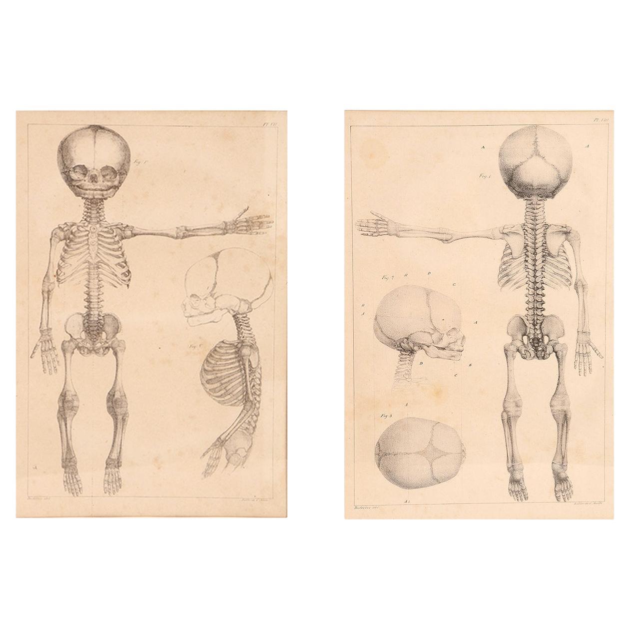
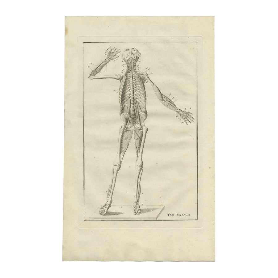
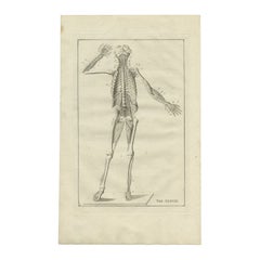
View AllAnatomy Print: Skeleton, Skull, Organs & Nerves Study – Antique 1867
Located in Langweer, NL
Anatomy Print: Skeleton, Skull, Organs & Nerves Study – Antique 1867
This detailed antique print presents a comprehensive anatomical study from the 19th century, depicting the human...
Category
Antique 1860s German Prints
Materials
Paper
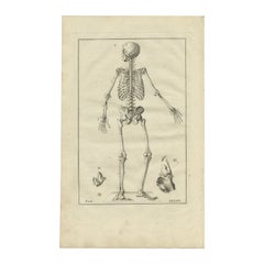
Decorative Antique Anatomy Print of the Human Skeleton, 1798
Located in Langweer, NL
Antique anatomy print of the human skeleton. This print originates from 'De Ontleedkundige Plaaten van B. Eustachius' published by J.B. Elwe.
Artists and Engravers: Bartolomeo Eus...
Category
Antique 18th Century Prints
Materials
Paper
$325 Sale Price
20% Off
Antique Anatomy Print of the Human Skeleton, 1798
Located in Langweer, NL
Antique anatomy print of the human skeleton. This print originates from 'De Ontleedkundige Plaaten van B. Eustachius' published by J.B. Elwe.
Artist...
Category
Antique 18th Century Prints
Materials
Paper
$326 Sale Price
20% Off
Antique Anatomy Print of the Human Skeleton, 1798
Located in Langweer, NL
Antique anatomy print of the human skeleton. This print originates from 'De Ontleedkundige Plaaten van B. Eustachius' published by J.B. Elwe.
...
Category
Antique 18th Century Prints
Materials
Paper
$326 Sale Price
20% Off
Antique Print of the Circulatory System and Nervous System, 1879
Located in Langweer, NL
Antique print titled 'Vue d'Ensemble de l'Appareil circulatoire et du système nerveux'. Color lithograph of the circulatory system and nervous system. This print originates from 'Le ...
Category
Antique 19th Century Prints
Materials
Paper
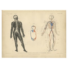
$153 Sale Price
20% Off
Antique Anatomical Print of the Nervous System in the Human Body '1843'
Located in Langweer, NL
Coloured anatomical plate (no. 17), showing the nervous system in the human body. From Jean-Baptiste Bourgery's 'Anatomie élementaire', published 1843, illustrated by Nicolas-Henri J...
Category
Antique Mid-19th Century Prints
Materials
Paper
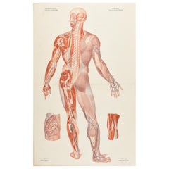
$1,080 Sale Price
25% Off
You May Also Like
Anatomical Print on Paper, Depicting a Fetus Skeleton, France, 19th Century
Located in Milan, IT
A rare couple of French anatomical lithographs of the fetus skeleton, in a vintage metal frame. Great quality and artistic composition.
Printe...
Category
Antique Early 19th Century French Prints
Materials
Metal

Original Vintage Medical Print-Skeleton, circa 1900
Located in St Annes, Lancashire
Great image of medical interest.
Unframed.
Published, circa 1900.
Category
Antique Early 1900s English Edwardian Prints
Materials
Paper
Human Anatomical Skeleton - Original Lithograph by Paul Gervais - 1854
By Paul Gervais
Located in Roma, IT
Human Anatomical Skeleton is an original lithograph on ivory-colored paper, realized by Paul Gervais (1816-1879). The artwork is from The Series of...
Category
1850s Modern Figurative Prints
Materials
Lithograph
$201 Sale Price
40% Off
Antique Anatomical Wall Chart Depicting the Human Skeleton
Located in Berghuelen, DE
Antique Anatomical Wall Chart Depicting the Human Skeleton
A rare 19th century anatomical wall chart depicting the human skeleton. On the sides On the...
Category
Early 20th Century German Victorian Prints
Materials
Paper
Antique Anatomical Chart Architecture of the Human Anatomy by E. Hoelemann
Located in Atlanta, GA
A great antique anatomical chart depicting the different sense organs. the chart is sheet number VI as part of the architecture of human anato...
Category
Antique Early 1900s Scandinavian Modern Posters
Materials
Paper
Anatomical drawing of a skeleton, made in pencil and ink, Italy 1890.
Located in Milan, IT
A painting containing an ancient anatomical pencil and ink drawing of a skeleton, created for the creation of anatomical tables. The antique frame, made of fir wood with rounded corn...
Category
Antique Late 19th Century Italian Paintings
Materials
Iron

More Ways To Browse
Heart Display
Eye Chart
Antique Human Skeleton
Medical Skull
Foo Dog Jar
Fornasetti Frame
Franco Albini Bar Cart
Frank Lloyd Wright Stained Glass
French Art Deco Tea Pot
French Commode Demilune
French Directoire Buffet
French Elm Commode
French Herbarium
French Kitchen Hutch
French Regency Bed
French Tea Trolley
Frog Wood Sculpture
George Iii Dressing Table